Catalogue

Rat anti Integrin alpha 6A / CD49f
Catalog number: MUB0908P$402.00
Add To Cart| Clone | NKI-GoH3 |
| Isotype | IgG2a |
| Product Type |
Monoclonal Antibody Primary Antibodies |
| Units | 0.1 mg |
| Host | Rat |
| Species Reactivity |
Human Mouse |
| Application |
Flow Cytometry Immunocytochemistry Immunohistochemistry (frozen) Immunoprecipitation |
Background
Integrins, are transmembrane glycoproteins that belong to the family of adhesion molecules. They promote interactions between cells and their environment being both other cells and the extra cellular matrix. Integrins are a family of heterodimeric membrane glycoproteins consisting of non-covalently associated subunits, being an α-subunit of 95 kDa that is conserved through the superfamily and a more variable β subunit of 150-170 kDa. More than 18 α and 8 β subunits with numerous splice variant isoforms have been identified in mammals. One of the integrin alpha subunits is the integrin alpha 6 (α6) subunit. Integrin α6 (CD49f) primarily associates with the integrin beta 1 (β1) or beta 4 (β4) subunit to form the α6β1 or laminin receptor and the α6β4 heterodimer. The GoH3 monoclonal antibody recognizes cell surface antigens on epithelial cells, endothelial cells and in a variety of tissues in both Human and Mouse, and reacts with glycoproteins Ic (α6 integrin, also known as CD49f) complexed with glycoprotein IIa (β1) as the VLA-6 (very late activation antigen) or laminin receptor on Human and Mouse platelets, lymphocytes, epithelial cells and a variety of other cell types. In normal epithelial cells and (colon) carcinoma cells, peripheral nerves, and endothelia however, glycoprotein Ic (α6) is not associated with IIa, but with a group of proteins collectively named β4. These β4 proteins can occur in multiple forms on certain cell types. The tumor associated homologue of this protein is the TSP-180 antigen, expressed in lung carcinomas, melanomas, Human tissue carcinomas and carcinoma cell lines but not on Human melanomas and fibroblasts. VLA-6 is expressed on peripheral T cells or thymocytes, the GoH3 monoclonal antibody induces a comitogenic signal and inhibits platelet adhesion to laminin, one of the three major components of the subendothelial matrix.
Source
GoH3 is a Rat monoclonal IgG2a antibody obtained by immunizing Sprague-Dawley Rats with Mouse (Balb/c) mammary tumor cells. The fusion partner was SP2/0 Mouse myeloma cells.
Product
Each vial contains 100 microliters of 1 mg/ml ProtG purified monoclonal antibody in PBS containing 0.09% sodium azide.
Specificity
The epitope recognized by GoH3 is loCated on glycoprotein Ic or the a6 subunit of integrin. This α6 (glycoprotein Ic) is expressed as a physical complex with glycoprotein IIa within the plasma membrane protein on platelets and with the b4 subunit of integrin on normal epithelial cells and (colon) carcinoma cells, peripheral nerves, and endohelia. The epitope of the monoclonal antibody is still available when the Ic glycoprotein is within the complex.
Applications
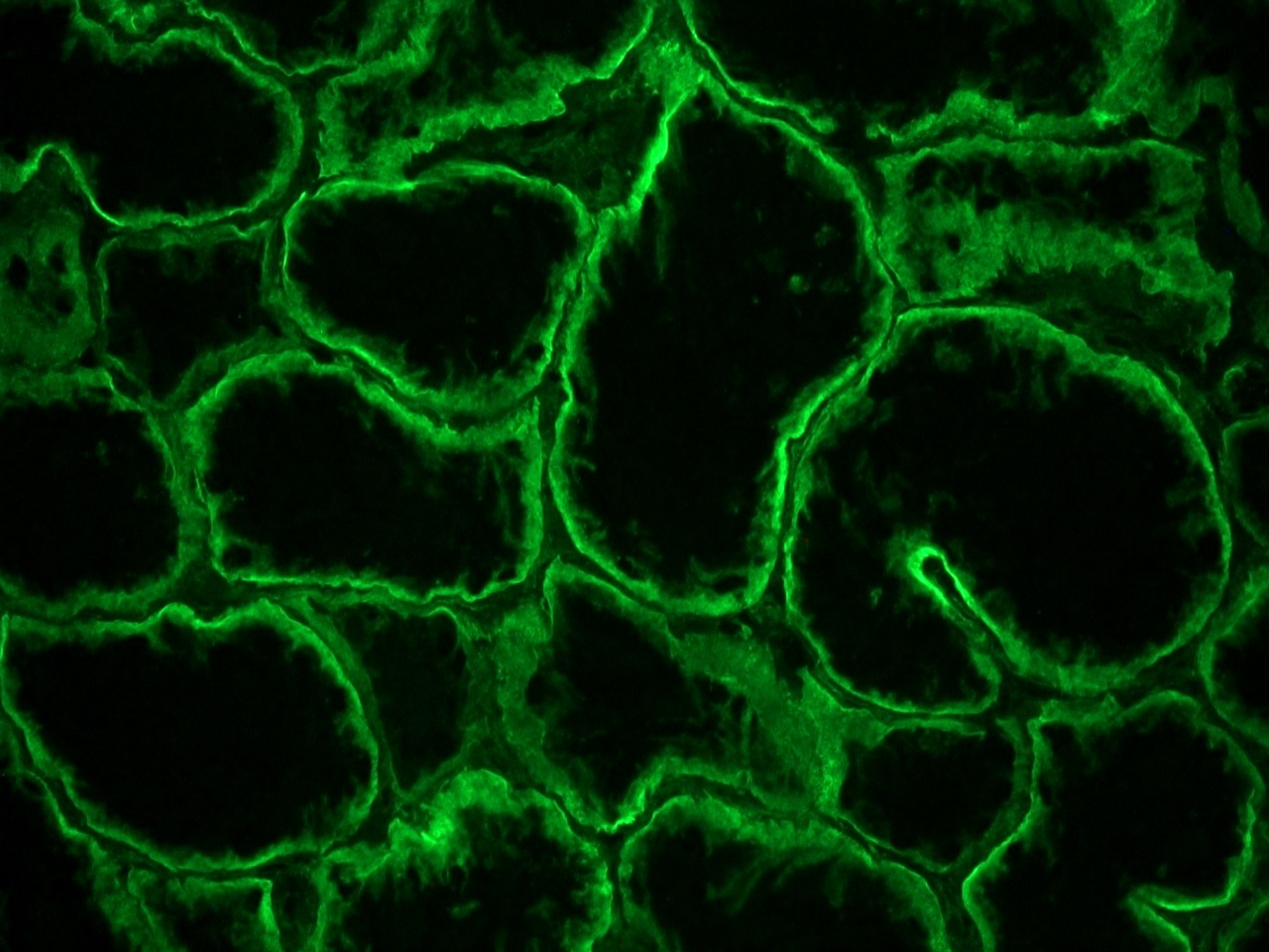
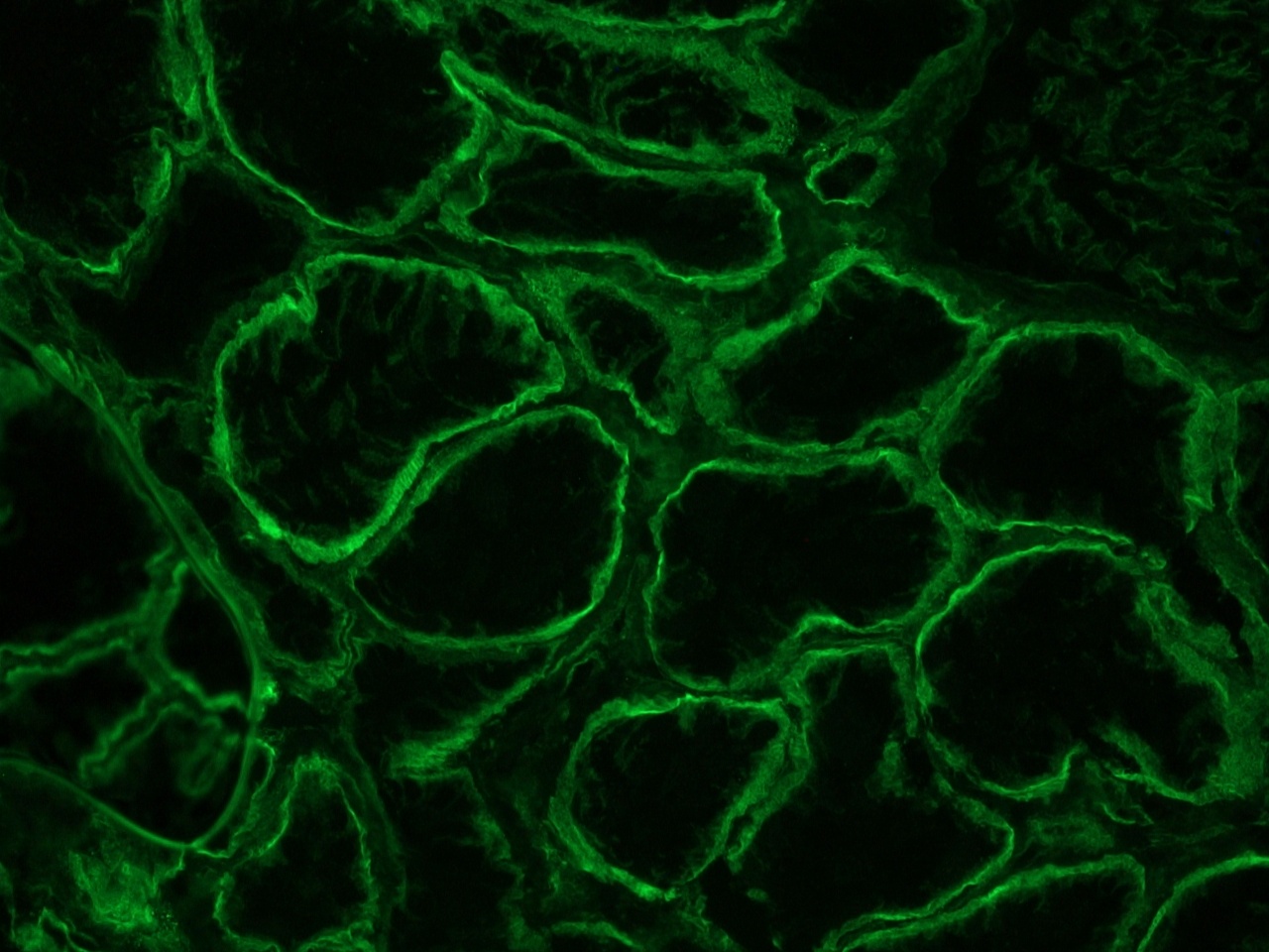
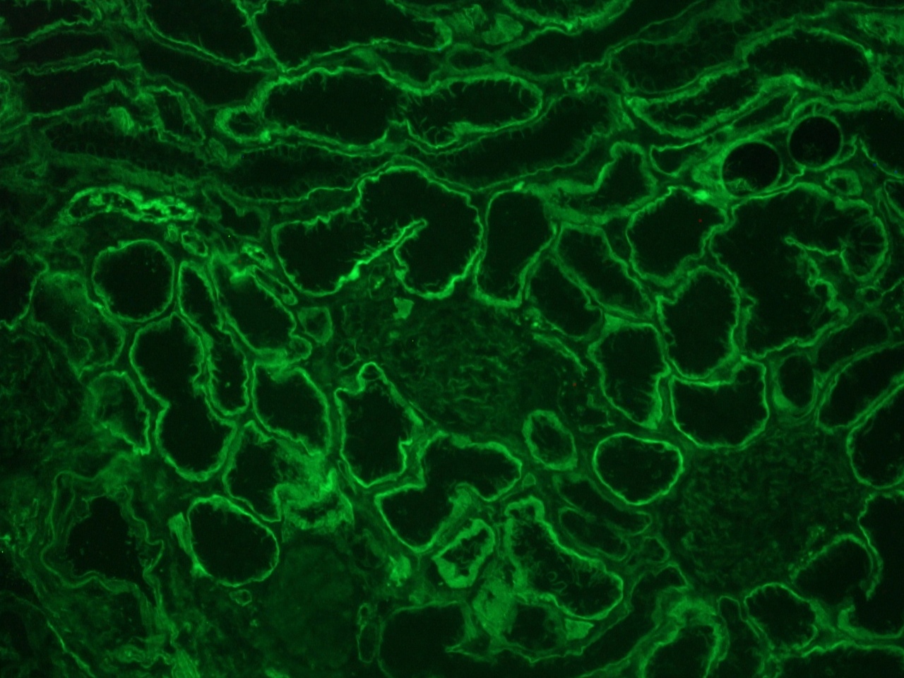
GoH3 can be used in flow cytometry, immunoprecipitation and immunocytochemistry. Optimal antibody dilution should be determined by titration. In immunohistochemistry staining of frozen Human tissues. GoH3 stains the basement membrane zone in skin sections, the myoepithelial cells in the mammary gland, tubules in kidney sections, acini and ducts in salivary glands, the epithelial layer in colonic crypts, the perineurium and the Schwann cells in sections of Human peripheral nerves. Smooth muscle cells, endothelial cells and striated muscle and the muscularis of the large intestine are not stained.
Storage
The antibody may be stored at +4°C. For prolonged storage prepare appropriate aliquots and store at or below -20°C. Prior to use, an aliquot is thawed slowly in the dark at ambient temperature, spun down again and used to prepare working dilutions by adding sterile phosphate buffered saline (PBS, pH 7.2). Repeated thawing and freezing should be avoided. Working dilutions should be stored at +4°C, not refrozen, and preferably used the same day. If a slight precipitation occurs upon storage, this should be removed by centrifugation. It will not affect the performance or the concentration of the product.
Shipping Conditions: The antibody is shipped at ambient temperature.
Caution
This product is intended FOR RESEARCH USE ONLY, and FOR TESTS IN VITRO, not for use in diagnostic or therapeutic procedures involving humans or animals. It may contain hazardous ingredients. Please refer to the Safety Data Sheets (SDS) for additional information and proper handling procedures. Dispose product remainders according to local regulations.This datasheet is as accurate as reasonably achievable, but our company accepts no liability for any inaccuracies or omissions in this information.
References
1. Ticchioni, M., Deckert, M., Bernard, G., Calandra, D., Breittmeyer, J.P., Imbert, V., Peyron, J.F. and Bernard, A. (1995). Comitogenic effects of very late activation antigens on CD3-stimulated Human thymocytes. J Immunol 154, 1207-15.
2. Sonnenberg, A., Janssen, H., Hogervorst, F., Calafat, J. and Hilgers, J. (1987). A complex of platelet glycoproteins Ic and IIa identified by a Rat monoclonal antibody. J Biol Chem 262, 10376-83.
3. Hemler, M.E., Crouse, C., Takada, Y. and Sonnenberg, A. (1988). Multiple very late antigen (VLA) heterodimers on platelets. J Biol Chem 263, 7660-5.
Safety Datasheet(s) for this product:
| EA_Sodium Azide |

Figure 2: Integrin alpha 6A immunostaining of the basolateral membrane of epithelial cells in a frozen section of human kidney using MUB0908P.

Figure 3: Integrin alpha 6A immunostaining of the basolateral membrane of epithelial cells in a frozen section of human kidney using MUB0908P.

Figure 4: Integrin alpha 6A immunostaining of the basolateral membrane of epithelial cells in a frozen section of human kidney using MUB0908P.
