Catalogue

Mouse anti Plakophilin-3
Catalog number: MUB1502P$402.00
Add To Cart| Clone | 12B11F8 |
| Isotype | IgG2a |
| Product Type |
Primary Antibodies |
| Units | 0.1 mg |
| Host | Mouse |
| Species Reactivity |
Canine Human Mouse Swine |
| Application |
Immunocytochemistry Immunohistochemistry (frozen) Immunoprecipitation Western Blotting |
Background
Plakophilin-3 is an Armadillo-like protein present in nuclei and desmosomes of epithelial cells. The expression pattern of this protein seems to be largely restricted to epithelial cell types. Plakophilin-3 can be detected along cell borders in a punctuate staining pattern typical for desmosomal proteins. In addition to the desmosomal immunolocalisation, immunostaining was observed as bright nuclear speckles. Thus, like plakophilin-1 and-2, plakophilin-3 displays a dual intracellular localisation in the desmosomal plaque and in the cell nucleus, and therefore is probably involved in signal transduction pathways between the plasma membrane and the nucleus. The human protein has a predicted molecular mass of 87 kD.
Source
12B11F8 is a mouse monoclonal IgG2a antibody derived by fusion of mouse myeloma cells with spleen cells from a mouse immunized with a synthetic peptide corresponding to the extreme carboxyterminal amino acid residues 779-793 (KLHRDFRAKGYRKED) of human plakophilin-3 coupled to keyhole limpet hemocyanin
Product
Each vial contains 100 ul 1 mg/ml purified monoclonal antibody in PBS containing 0.09% sodium azide.
Formulation: Each vial contains 100 ul 1 mg/ml purified monoclonal antibody in PBS containing 0.09% sodium azide.
Specificity
12B11F8 reacts with an epitope loCated between residues 779-793 in Human plakophilin-3.
Applications
12B11F8 is suitable for immunocytochemistry (methanol-fixed cells), immunohistochemistry on frozen tissues, immunoprecipitation and Western blot detection. For frozen tissues use a PBS buffer containing 0.1 mM CaCl2 and 0.1 mM MgCl2. For optimal results, fixed cells or tissues should be treated with 0.2% Triton-X 100 for 15 min. prior to incubation with primary antibody. Optimal antibody dilution should be determined by titration; recommended range is 1:25 – 1:100 for immunohistochemistry with avidin-biotinylated horseradish peroxidase complex (ABC) as detection reagent, and 1:100 – 1:1000 for immunoblotting applications.
Storage
The antibody is shipped at ambient temperature and may be stored at +4°C. For prolonged storage prepare appropriate aliquots and store at or below -20°C. Prior to use, an aliquot is thawed slowly in the dark at ambient temperature, spun down again and used to prepare working dilutions by adding sterile phosphate buffered saline (PBS, pH 7.2). Repeated thawing and freezing should be avoided. Working dilutions should be stored at +4°C, not refrozen, and preferably used the same day. If a slight precipitation occurs upon storage, this should be removed by centrifugation. It will not affect the performance or the concentration of the product.
Caution
This product is intended FOR RESEARCH USE ONLY, and FOR TESTS IN VITRO, not for use in diagnostic or therapeutic procedures involving humans or animals. It may contain hazardous ingredients. Please refer to the Safety Data Sheets (SDS) for additional information and proper handling procedures. Dispose product remainders according to local regulations.This datasheet is as accurate as reasonably achievable, but our company accepts no liability for any inaccuracies or omissions in this information.
References
1. Bonné, S., van Hengel, J., Nollet, F., Kools, P., and van Roy, F. (1999). Plakophilin-3, a novel armadillo-like protein present in nuclei and desmosomes of epithelial cells. J Cell Sci 112, 2265-2276.
2. Bonné, S., Gilbert, B., Hatzfeld, M., Chen, X., Green, K.J., van Roy, F. (2003). Defining desmosomal plakophilin-3 interactions. J Cell Biol 161,2, 403-416.
Safety Datasheet(s) for this product:
| EA_Sodium Azide |

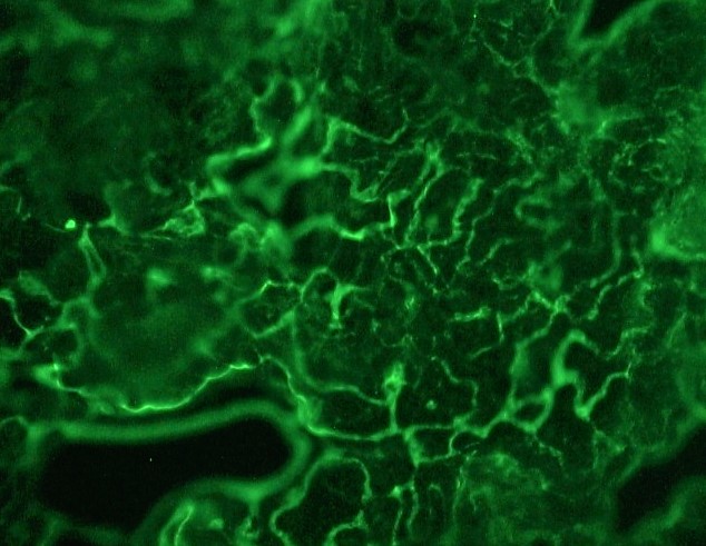
Figure 1. Indirect immunohistochemical staining of MUB1502P (clone 12B11F8) on a frozen tissue section of dog skin, showing the specific desmosomal localization of plakophilin-3. Antibody dilution 1:25.

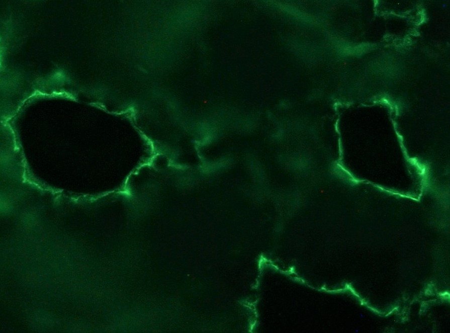
Figure 2. Indirect immunohistochemical staining of MUB1502P (clone 12B11F8) on a frozen tissue section of dog skin, showing the specific desmosomal localization of plakophilin-3. Antibody dilution 1:50.

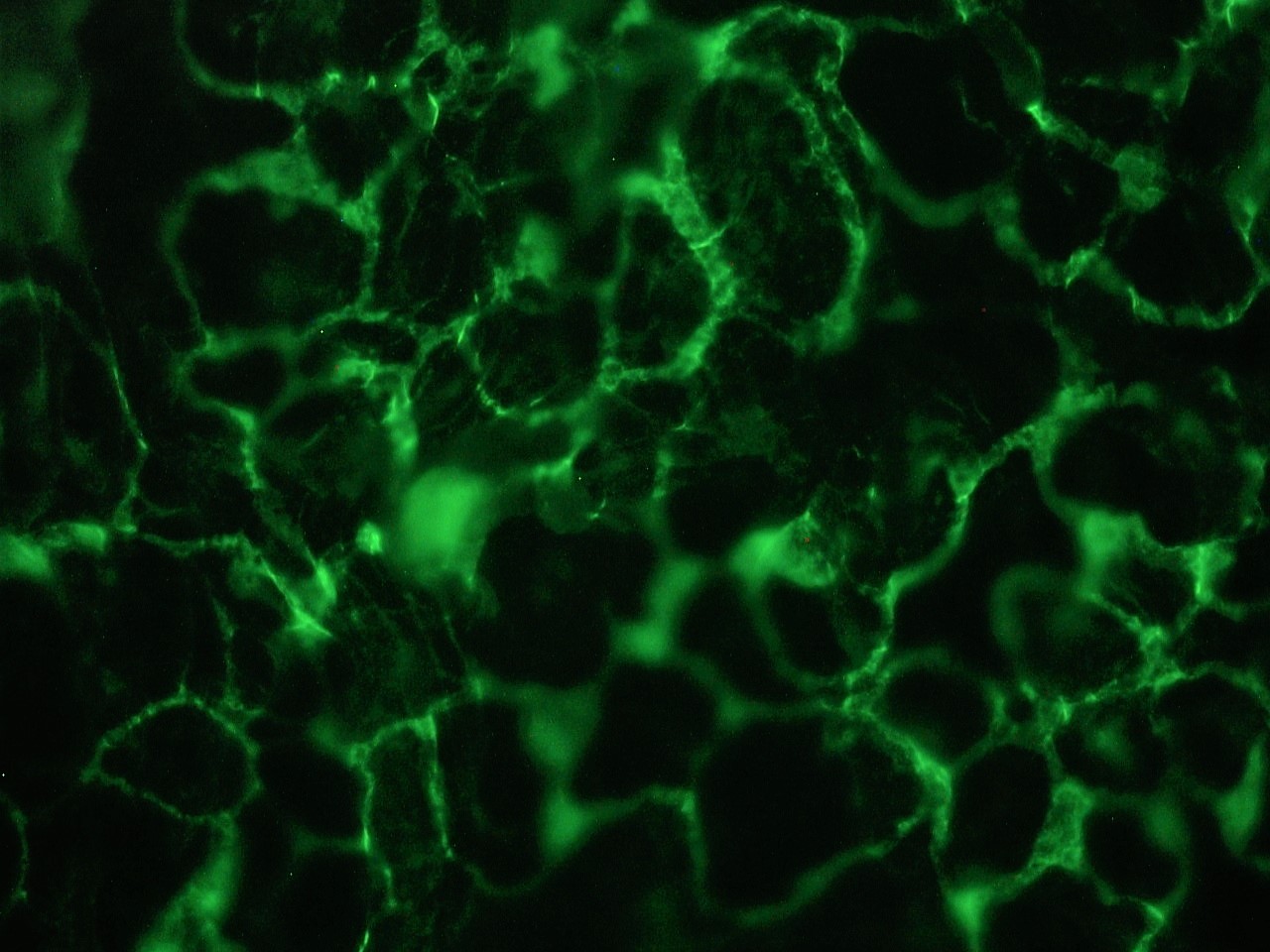
Figure 3. Indirect immunohistochemical staining of MUB1502P (clone 12B11F8) on a frozen tissue section of dog skin, showing the specific desmosomal localization of plakophilin-3. Antibody dilution 1:100.
