Mouse anti Luciferase (Firefly) Protein Luciferase (N-Terminus)
Catalog number: X1171M$335.00
Add To Cart| Clone | Luci17 |
| Isotype | IgG1 |
| Product Type |
Monoclonal Antibody |
| Units | 100 µg |
| Host | Mouse |
| Application |
Immunohistochemistry Western Blotting |
Background
Analysis of gene expression is most commonly assayed by transient transfection. Systems are generally based on the use of fusion genes which are inserted into cells, and the gene expression is assayed within 48 hours after introduction of DNA. Usually the fusion consists of the promoter binding site or enhancer sequence under study which is attached to a reporter gene. The amount of the reporter protein synthesized under the experimental conditions, is presumed to reflect the ability of the sequences studied to direct or promote transcription. Several enzymes are commonly used as reporter proteins, among them are chloramphenicol acetyl transferase (CAT), -galactosidase, human growth hormone (hGH) and luciferase. Luciferase has become one of the widely used reporter enzymes. The enzyme catalyzes a bioluminescent reaction which requires the substrate luciferin as well as Mg+2 and ATP. Mixing these reagents with the cell extract containing luciferase, results in a flash of light that decays rapidly. This light can be detected by a luminometer. The total light emission is proportional to the luciferase activity of the sample. The use of an antibody to detect luciferase can provide an alternative detection assay which directly detects luciferase protein levels, and thus has the advantage that it does not require luciferase activity and is not dependent on rapid kinetics. Moreover, antibodies can detect the luciferase enzyme expression in situ, providing a means to study the localized signal sequences using luciferase as a reporter gene. Reacts with Luciferase (Firefly) Protein.
Source
Immunogen: Hybridoma produced by the fusion of splenocytes from mice immunized with luciferase protein isolated from Photinus pyralis.
Product
Product Form: Unconjugated
Formulation: Provided as solution in phosphate buffered saline with 0.08% sodium azide
Purification Method: Protein A/G Chromatography
Concentration: See vial for concentration
Applications
Detects luciferase protein by Western blot in C. elegans and Drosophilia melanogaster tissues, human fibroblast, mouse macrophage, kidney, liver and cortex as well as NIH3T3, Jurkat and BHR21 cell lines. Detects luciferase with little to no background signal. Optimal concentration should be evaluated by serial dilutions.
Functional Analysis: Western Blotting
Positive Control: Purified luciferase protein
Storage
Product should be stored at -20°C. Aliquot to avoid freeze/thaw cycles
Product Stability: See expiration date on vial
Shipping Conditions: Ship at ambient temperature, freeze upon arrival
Caution
This product is intended FOR RESEARCH USE ONLY, and FOR TESTS IN VITRO, not for use in diagnostic or therapeutic procedures involving humans or animals. It may contain hazardous ingredients. Please refer to the Safety Data Sheets (SDS) for additional information and proper handling procedures. Dispose product remainders according to local regulations.This datasheet is as accurate as reasonably achievable, but our company accepts no liability for any inaccuracies or omissions in this information.
References
1. Aoki, Y., et al. Selective stimulation of G-CSF gene expression in macrophages by a stimulatory monoclonal antibody as detected by a luciferase reporter gene assay. J. Leukoc. Biol. 2000, 68, 757-764
2. Nicolas, M.T., et al. Immunogold labeling of luciferase in the luminous bacterium Vibrio Harveyi after fast-freeze fixation and different freeze-substitution and embedding procedures. J. Histochem. Cytochem. 1989, 37, 663-674
Safety Datasheet(s) for this product:
| EA_Sodium Azide |

|
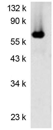
" Western blot analysis using Luciferase antibody (Cat. No. X1171M) on recombinant luciferase protein." |

" Western blot analysis using Luciferase antibody (Cat. No. X1171M) on recombinant luciferase protein."
