Catalogue

Mouse anti Human MyoD1
Catalog number: X1414M$465.00
Add To Cart| Clone | 5.2F |
| Isotype | IgG2a |
| Product Type |
Monoclonal Antibody |
| Units | 100 µg |
| Host | Mouse |
| Species Reactivity |
Chicken Human Mouse Pig Rat |
| Application |
ELISA Immunofluorescense Immunohistochemistry (frozen & paraffin) Immunoprecipitation Western Blotting |
Background
X1414M monoclonal antibody, clone 5.2F recognizes a protein of 45kDa, identified as MyoD1. The epitope of this antibody maps in between amino acid 3-56 in the N-terminus of mouse MyoD1 protein. It does not cross react with myogenin, Myf5, or Myf6. Occassionally nuclear expression/staining of MyoD1 is seen in ectomesenchymoma and a subset of Wilms tumors. Weak cytoplasmic staining/presence is observed in several non-muscle tissues, including glandular epithelium and also in rhabdomyosarcomas, neuroblastomas, Ewings sarcomas and alveolar soft part sarcomas. The 5.2F antibody to MyoD1 labels the nuclei of myoblasts in developing muscle tissues. MyoD1 is not detected in normal adult tissue.
Synonyms: Myoblast determination protein 1, Myogenic factor 3, MyoD1, Myf-3
Source
Hybridoma produced by the fusion of splenocytes from BALB/c mice immunized with recombinant mouse MyoD1 protein and mouse myeloma Sp2/0-Ag14 cells.
Immunogen: Recombinant mouse MyoD1 protein.
Product
Purified monoclonal antibody 5.2F in phosphate buffered saline with 0.08% sodium azide
Product Form: Unconjugated
Purification Method: Protein A/G Chromatography
Concentration: See vial for concentration
Applications
This antibody can be used for electron microscopy, ELISA, immunofluorescence, immunoprecipitation (2 µg/mg of protein lysate), Western blotting (1 µg/ml) and immunohistochemistry on frozen and formalin/paraffin fixed tissues (2-4 µg/ml). These working concentrations are merely an indication. Optimal working concentrations should, however, be evaluated by serial dilutions by the customer.
Functional Analysis: Western Blotting
Positive Control: Rhabdomyosarcoma, SW80 cells
Storage
Product should be stored at -20°C. Aliquot to avoid freeze/thaw cycles
Product Stability: See expiration date on vial
Shipping Conditions: Ship at ambient temperature, freeze upon arrival
Caution
This product is intended FOR RESEARCH USE ONLY, and FOR TESTS IN VITRO, not for use in diagnostic or therapeutic procedures involving humans or animals. It may contain hazardous ingredients. Please refer to the Safety Data Sheets (SDS) for additional information and proper handling procedures. Dispose product remainders according to local regulations.This datasheet is as accurate as reasonably achievable, but our company accepts no liability for any inaccuracies or omissions in this information.
References
1. Thulasi R; et al. Alpha 2a-interferon-induced differentiation of human alveolar rhabdomyosarcoma cells: correlation with down-regulation of the insulin-like growth factor type I receptor. Cell Growth and Differentiation, 1996 Apr, 7(4):531-41
2. Wesche WA; et al. Immunohistochemistry of MyoD1 in adult pleomorphic soft tissue sarcomas. American Journal of Surgical Pathology, 1995, 19(3):261-9.
3. Parham DM; et al. Immunohistochemical analysis of the distribution of MyoD1 in muscle biopsies of primary myopathies and neurogenic atrophy. Acta Neuropathologica, 1994, 87:605-11.
4. Tallini G; et al. Myogenic regulatory protein expression in adult soft tissue sarcomas. A sensitive and specific marker of skeletal muscle differentiation. American Journal of Pathology, 1994 Apr, 144(4):693-701.
5. Dias P; et al. Monoclonal antibodies to the myogenic regulatory protein MyoD1: epitope mapping and diagnostic utility. Cancer Research, 1992 Dec 1, 52(23):6431-9.
6. Rosai J; et al. MyoD1 protein expression in alveolar soft part sarcoma as confirmatory evidence of its skeletal muscle nature. American Journal of Surgical Pathology, 1991 Oct, 15(10):974-81.
7. Kolm PJ and Sive HL. Efficient hormone-inducible proeten function in Xenopus laevis. Developmental Biology, 1995, 171:267-272.
8. Attia GM, Atef H, Elmansy RA. Autologus platelet rich plasma enhances satellite cells expression of MyoD and exerts angiogenic and antifibrotic effects in experimental rat model of traumatic skeletal muscle injury. Egyptian Journal of Histology, 2017, 40: 443-458.
9.Miersch C, Stange K and Roentgen M. Separation of functionally divergent muscle precursor cell populations from porcine juvenile muscles by discontinuous Percoll density gradient centrifugation. BMC Cell Biology, 2018, 19:2
Protein Reference(s)
Database Name: UniProt
Accession Number: P15172 (Human) P10085 (Mouse) Q02346 (Rat) P16075
Species Accession: Human, Rat, Mouse, Chicken
Safety Datasheet(s) for this product:
| EA_Sodium Azide |
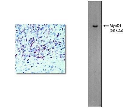
"Left: Immunohistochemical staining using MyoD1 antibody on formalin fixed, paraffin embedded human rhabdomyosarcoma. Right: Western blot using 60 ng of purified MyoD1 protein with MyoD1 antibody at 2 µg/ml and detected using goat anti-mouse HRP (Cat. No. X1208P) and visualized with Pierce West-Femto substrate."
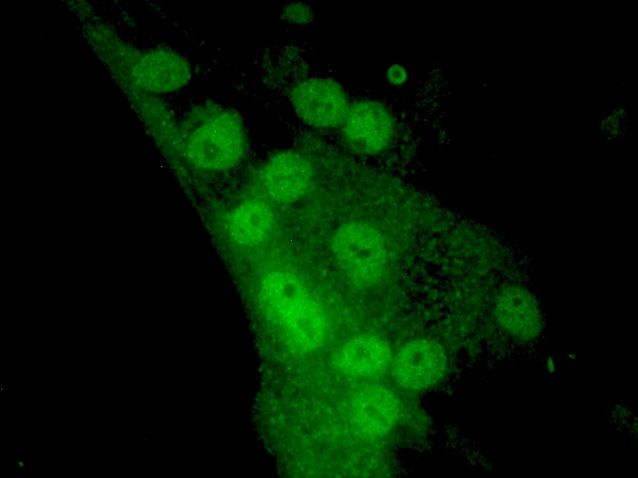
Figure 2. MyoD1 immunostaining of nuclei in MyoD1 transfected cells induced to undergo muscle cell differentiation.
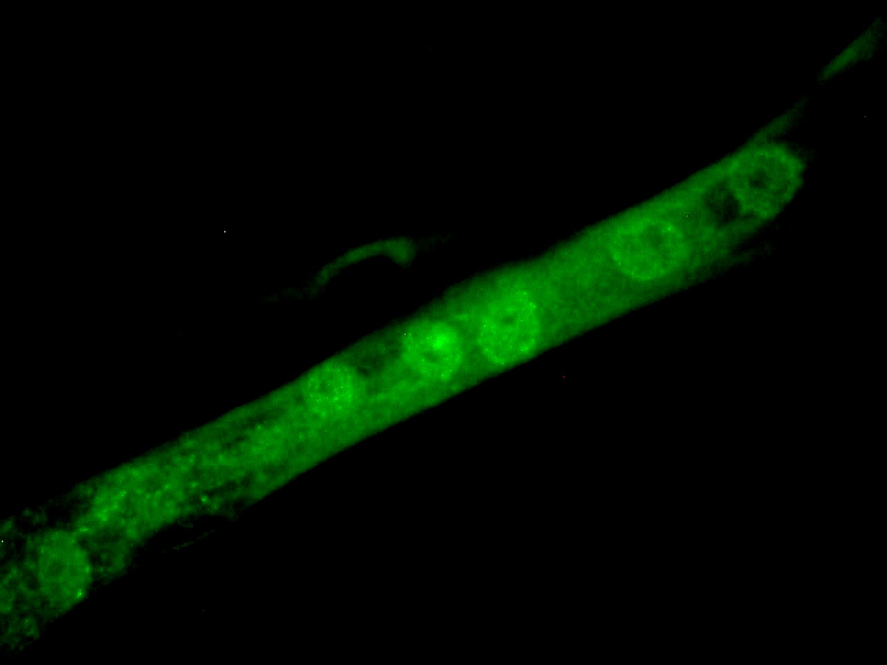
Figure 3. MyoD1 immunostaining of nuclei in MyoD1 transfected cells induced to undergo muscle cell differentiation.
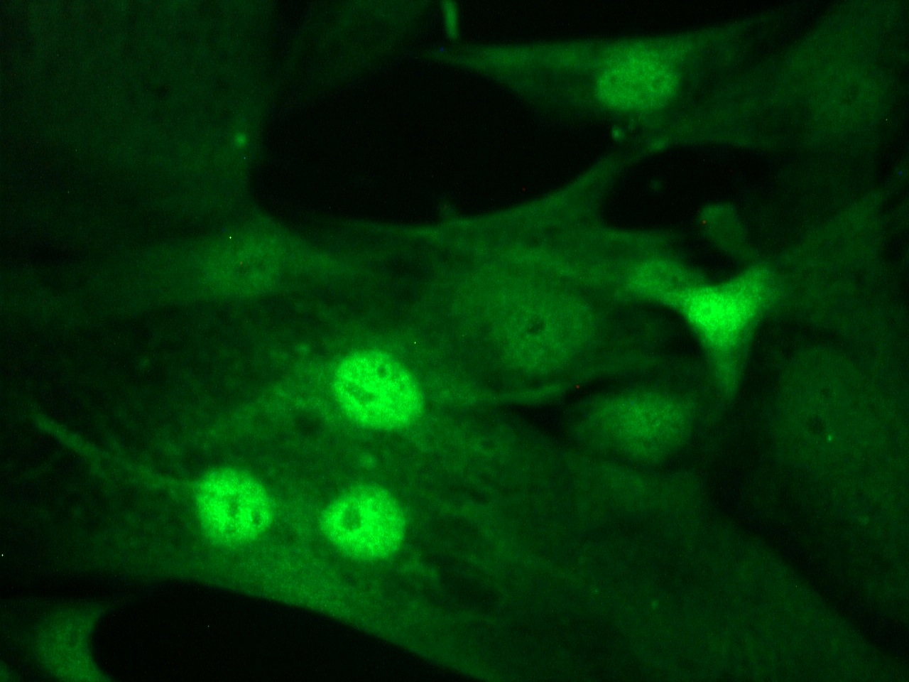
Figure 4. MyoD1 immunostaining of nuclei in MyoD1 transfected cells induced to undergo muscle cell differentiation.
