Catalogue

Mouse anti Human Keratin (4,5,6,8,10,13,18)
Catalog number: X1260M$335.00
Add To Cart| Clone | C11 |
| Isotype | IgG1 |
| Product Type |
Monoclonal Antibody |
| Units | 100 µg |
| Host | Mouse |
| Species Reactivity |
Human |
| Application |
Immunohistochemistry (frozen & paraffin) Western Blotting |
Background
Cytokeratins (CK) are intermediate filaments of epithelial cells, both in keratinizing tissue (ie., skin) and non-keratinizing cells (ie., mesothelial cells). Although not a traditional marker for endothelial cells, cytokeratins have also been found in some microvascular endothelial cells. At least 20 different cytokeratins in the molecular range of 40-70 kDa and isoelectric points of 5-8.5 can be identified using two dimensional gel electrophoresis. Biochemically, most members of the CK family fall into one of two classes, type I (acidic polypeptides) and type II (basic polypeptides). At least one member of the acidic family and one member of the basic family is expressed in all epithelial cells. Monoclonal antibodies to cytokeratin proteins can be useful markers for tumor identification and classification.
Source
Hybridoma produced by the fusion of splenocytes from mice immunized with cytoskeleton preparation from human A431 carcinoma cells and mouse myeloma cells.
Product
Product Form: Unconjugated
Formulation: Provided as solution in phosphate buffered saline with 0.08% sodium azide
Purification Method: Purified by saturated ammonium sulphate precipitation
Concentration: See vial for concentration
Applications
X1260M detects keratins 4,5,6,8,10,13 & 18 by Western blot. Immunohistochemistry can be performed on frozen tissues and paraffin sections. Optimal concentration should be evaluated by serial dilutions.
X1260M reacts with a variety of normal and neoplastic epithelia. Reacting with simple epithelium and both basal and superbasal layers of cornifying and non-cornifying squamous epithelium this antibody is also useful in staining cultured epithelial cell lines. It is useful in differentiating epithelial tumors from non-epithelial tumors.
Functional Analysis: Western blotting
Storage
Product should be stored at -20°C. Aliquot to avoid freeze/thaw cycles
Product Stability: See expiration date on vial
Shipping Conditions: Ship at ambient temperature, freeze upon arrival
Caution
This product is intended FOR RESEARCH USE ONLY, and FOR TESTS IN VITRO, not for use in diagnostic or therapeutic procedures involving humans or animals. It may contain hazardous ingredients. Please refer to the Safety Data Sheets (SDS) for additional information and proper handling procedures. Dispose product remainders according to local regulations.This datasheet is as accurate as reasonably achievable, but our company accepts no liability for any inaccuracies or omissions in this information.
References
1. Folia Biol. 1989, 35, 373-382
2. Neoplasma 1990, 37, 333-342
3. J. Tumor Marker Oncol. 1990, 5, 219
4. Int. J. Cancer 1988, Suppl. 3, 50-55
Protein Reference(s)
Database Name: SwissProt
Accession Number: P13647
Species Accession: Human
Safety Datasheet(s) for this product:
| EA_Sodium Azide |

|
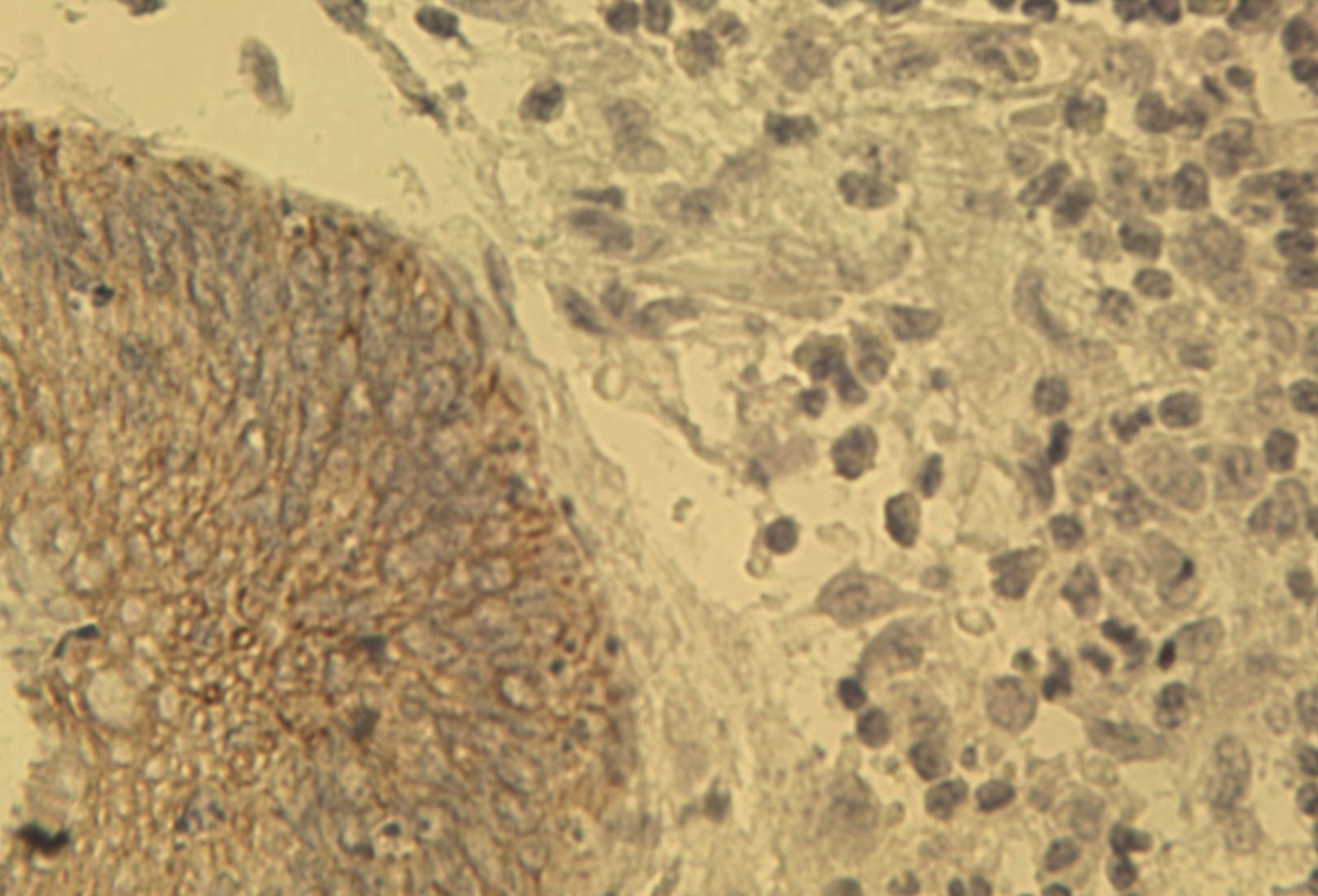
Figure 1. Immunohistochemical staining of human colon cancer tissue using pan Keratin antibody (Cat. No. X1260M) at 2.5 μg/ml. |

Figure 1. Immunohistochemical staining of human colon cancer tissue using pan Keratin antibody (Cat. No. X1260M) at 2.5 μg/ml.
