Cancer is an incredibly complex disease involving the aberrant expression and function of many different proteins. p53 is one of the most widely studied examples, with approximately 50% of human cancers known to carry mutations in the gene (TP53) encoding this essential nuclear transcription factor1.
Under normal conditions, p53 works as a tumor suppressor to enable cells to respond to DNA damage. Where DNA damage is mild, p53 mediates cell cycle arrest to allow repairs to be carried out before cells continue cycling; where damage is more severe, p53 instead promotes apoptosis to prevent faulty DNA being passed on to daughter cells2.
The tumor suppressing function of p53 is inactivated in many human cancers, including those of the breast, colon and lung. Inactivation occurs not only as of the result of TP53 mutations, but also due to various oncogenic events known to decrease the activity of the wild type p53 protein. For example, overexpression of MDM2 – the primary negative regulatory factor of p53 – leads to p53 being degraded by the proteasome3.
Researchers studying p53 to better understand its role in the onset and progression of cancer rely on high-quality anti-p53 antibodies. DO-1 is one of the most well-cited clones – something that is perhaps unsurprising since DO-1 was first described in the literature almost 30 years ago4. After initially being integrated into an ELISA for evaluating the p53 status of breast biopsy material, DO-1 has since become a routinely used tool within pathology to examine p53 protein expression in human tissues5,6.
High-quality antibodies for p53 research
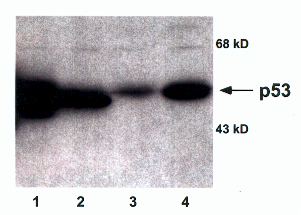
We offer numerous products to advance p53 research, including the mouse monoclonal anti-p53 antibody, DO-1 (MUB1501P). This has been epitope-mapped to the N-terminus (amino acids 11-25) of p53 and validated for Western blotting, ELISA, flow cytometry and immunocytochemistry, as well as for IHC with both frozen and paraffin-embedded tissue samples.
Complementing DO-1, our product portfolio also includes other anti-p53 antibodies raised in mouse hosts – such as clone X77 that epitope maps to amino acids 16-25 (X1157M). We also offer anti-p53 antibodies raised in alternative host species, like our sheep anti-human p53 antibody (A145P); these can be used to increase the diversity of multiplexed antibody panels, providing researchers with greater experimental flexibility.
- How does p53 induce apoptosis and how does this relate to p53-mediated tumour suppression? Aubrey BJ et al, Cell Death Differ. 2018 Jan;25(1):104-113
- Role of p53 in Cell Death and Human Cancers, Ozaki T and Nakagawara A, Cancers (Basel) 2011 Mar 3;3(1):994-1013
- The role of MDM2 amplification and overexpression in therapeutic resistance of malignant tumors, Hou H et al, Cancer Cell Int. 2019 Aug 22;19:216
- An immunochemical analysis of the human nuclear phosphoprotein p53. New monoclonal antibodies and epitope mapping using recombinant p53, Vojtĕsek B et al, J Immunol Methods
- . 1992 Jul 6;151(1-2):237-44
- Comparison between p53 staining in tissue sections and p53 proteins levels measured by an ELISA technique, Vojtĕsek B et al, Br J Cancer. 1993 Jun;67(6):1254-8
- Understanding p53 functions through p53 antibodies, Sabapathy K and Lane DP, J Mol Cell Biol. 2019 Apr 1;11(4):317-329