Catalogue

Mouse anti Bromodeoxyuridine / BrdU
Catalog number: MUB0200P$402.00
Add To Cart| Clone | IIB5 |
| Isotype | IgG1 |
| Product Type |
Primary Antibodies |
| Units | 0.1 mg |
| Host | Mouse |
| Application |
Flow Cytometry Immunocytochemistry Immunohistochemistry (frozen) Immunohistochemistry (paraffin) |
Background
The immunocytochemical detection of bromodeoxyuridine (BrdU) incorporated into DNA is a powerful tool to study the cytokinetics of normal and neoplastic cells. In vitro or in vivo labeling of tumor cells with the thymidine analogue BrdU and the subsequent detection of incorporated BrdU with specific anti-BrdU monoclonal antibodies is an accurate and comprehensive method to quantitate the degree of DNA-synthesis. BrdU is incorporated into the newly synthezised DNA of the S-phase cells and can thus provide an estimate for the fraction of cells in S-phase. Also dynamic proliferative information (such as the S-phase transit Rate and the potential doubling time) can be obtained, by means of bivariate BrdU/DNA flow cytometric analysis.
Source
IIB5 is a Mouse monoclonal IgG1 antibody derived by fusion of SP2/0-Ag14 Mouse myeloma cells with spleen cells from a BALB/c Mouse intraperitoneally immunized with BrdU conjugated to Bovine Serum albumin.
Immunogen: BrdU conjugated to Bovine Serum Albumin
Product
Each vial contains 100 ul 1 mg/ml purified monoclonal antibody in PBS containing 0.09% sodium azide.
Formulation: Each vial contains 100 ul 1 mg/ml purified monoclonal antibody in PBS containing 0.09% sodium azide.
Purification Method: ProtG affinity chromatography
Concentration: 1 mg/mL
Specificity
IIB5 reacts with bromodeoxyuridine (BrdU) also when incorporated into nuclear DNA.The antibody is known to cross-react with Iododeoxyuridine (IdU). Although we have no specific information concerning chlorodeoxyuridine (CldU), it is to be expected that also this antigen is recognized by IIB5.
Applications
IIB5 is suitable for flow cytometry and immunohistochemistry on frozen and paraffin-embedded tissues. It can also be applied in immunocytochemical staining procedures on cultured cells. Optimal antibody dilution should be determined by titration; recommended range is 1:100 – 1:200 for flow cytometry, and for immunohistochemistry/immunocytochemistry with avidin-biotinylated horseradish peroxidase complex (ABC) as detection reagent.
Storage
The antibody is shipped at ambient temperature and may be stored at +4°C. For prolonged storage prepare appropriate aliquots and store at or below -20°C. Prior to use, an aliquot is thawed slowly in the dark at ambient temperature, spun down again and used to prepare working dilutions by adding sterile phosphate buffered saline (PBS, pH 7.2). Repeated thawing and freezing should be avoided. Working dilutions should be stored at +4°C, not refrozen, and preferably used the same day. If a slight precipitation occurs upon storage, this should be removed by centrifugation. It will not affect the performance or the concentration of the product.
Shipping Conditions: Ship at ambient temperature.
Caution
This product is intended FOR RESEARCH USE ONLY, and FOR TESTS IN VITRO, not for use in diagnostic or therapeutic procedures involving humans or animals. It may contain hazardous ingredients. Please refer to the Safety Data Sheets (SDS) for additional information and proper handling procedures. Dispose product remainders according to local regulations.This datasheet is as accurate as reasonably achievable, but our company accepts no liability for any inaccuracies or omissions in this information.
References
1. Schutte, B., Reynders, M. M., van Assche, C. L., Hupperets, P. S., Bosman, F. T., and Blijham, G. H. (1987). An improved method for the immunocytochemical detection of bromodeoxyuridine labeled nuclei using flow cytometry, Cytometry 8, 372-6.
2.
2. Tinnemans, M. M., Schutte, B., Lenders, M. H., Ten Velde, G. P., Ramaekers, F. C., and Blijham, G. H. (1993). Cytokinetic analysis of lung cancer by in vivo bromodeoxyuridine labelling, Br J Cancer 67, 1217-22.
3.
3. Schutte, B., Tinnemans M.M.F.J., Pijpers, G.F.P., Lenders, M.J.H. and Ramaekers, F. (1995). Three parameter flow cytometric analysis for simultaneous detection of cytokeratin, proliferation associated antigens and DNA content. Cytometry 21, 177-86.
4.
4. van Engeland, M., Kuijpers, H. J., Ramaekers, F. C., Reutelingsperger, C. P., and Schutte, B. (1997). Plasma membrane alterations and cytoskeletal changes in apoptosis. Exp Cell Res 235, 421-430.
5.
5. Schutte, B., Nieland, L., van Engeland, M., Henfling, M. E., Meijer, L., and Ramaekers, F. C. (1997). The effect of the cyclin-dependent kinase inhibitor olomoucine on cell cycle kinetics. Exp Cell Res 236, 4-15.
Safety Datasheet(s) for this product:
| EA_Sodium Azide |
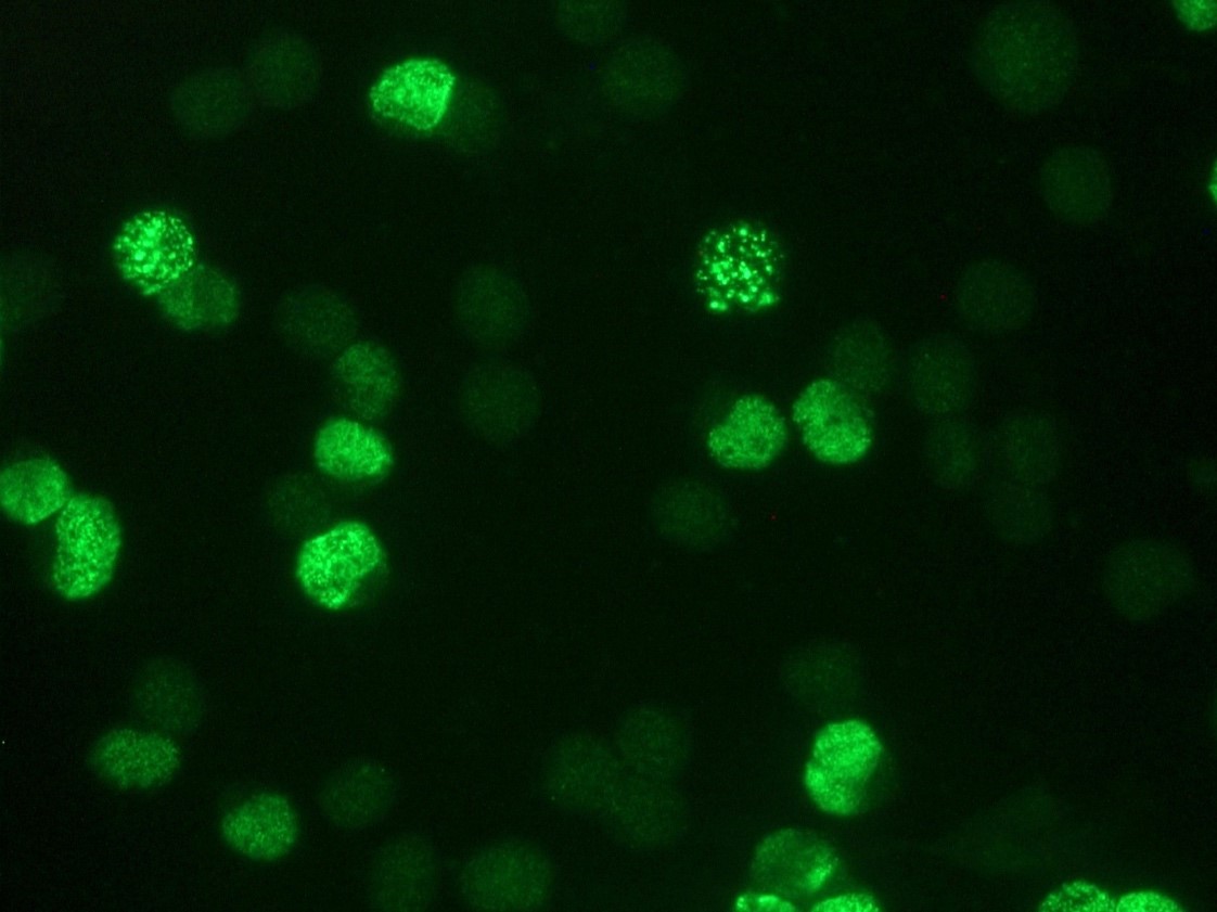
Figure 1a: Flow cytometric analysis of the BrdU-labeled fraction in a tissue cell culture.
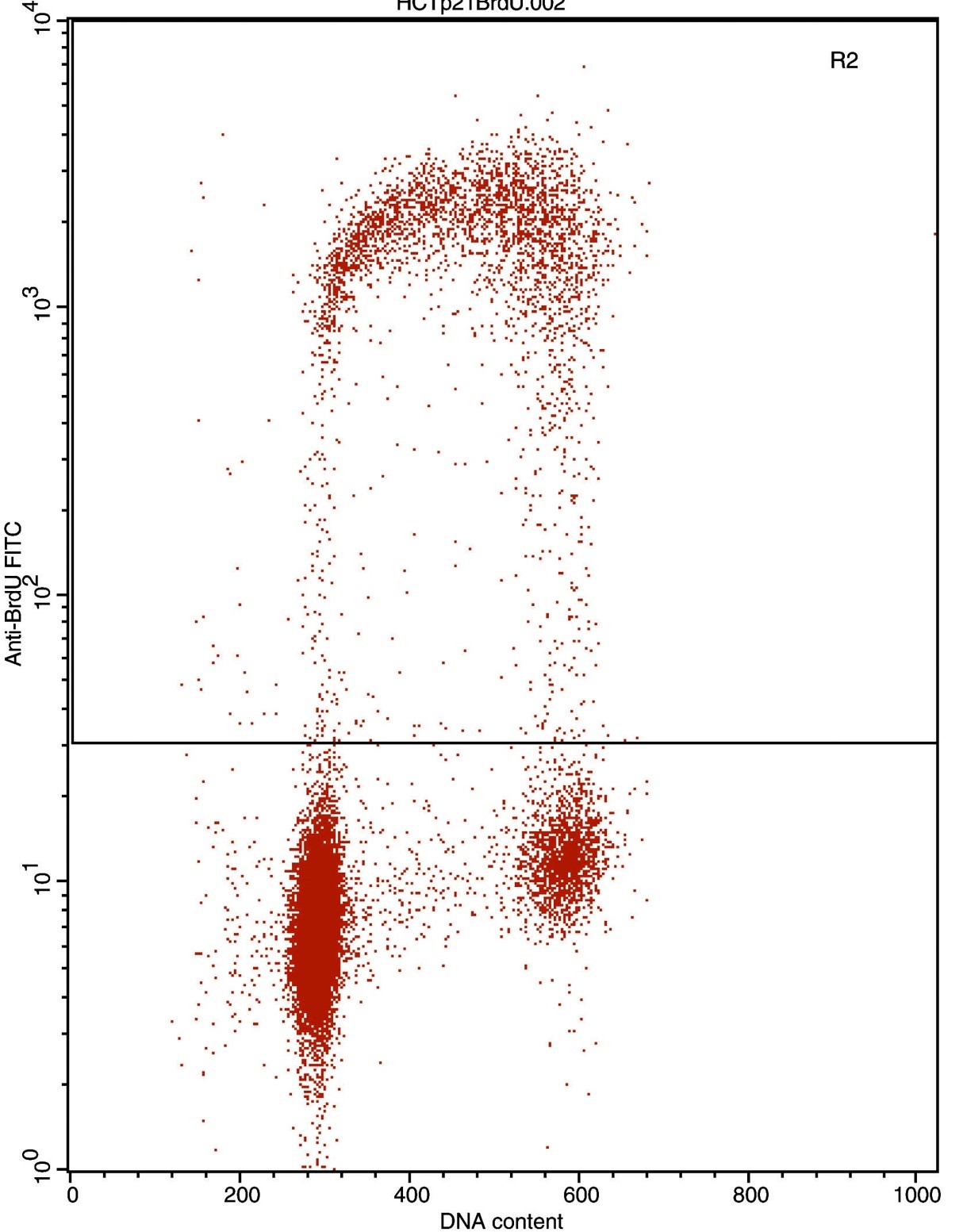
Figure 1b: Flow cytometric analysis of the BrdU-labeled fraction in a tissue cell culture.
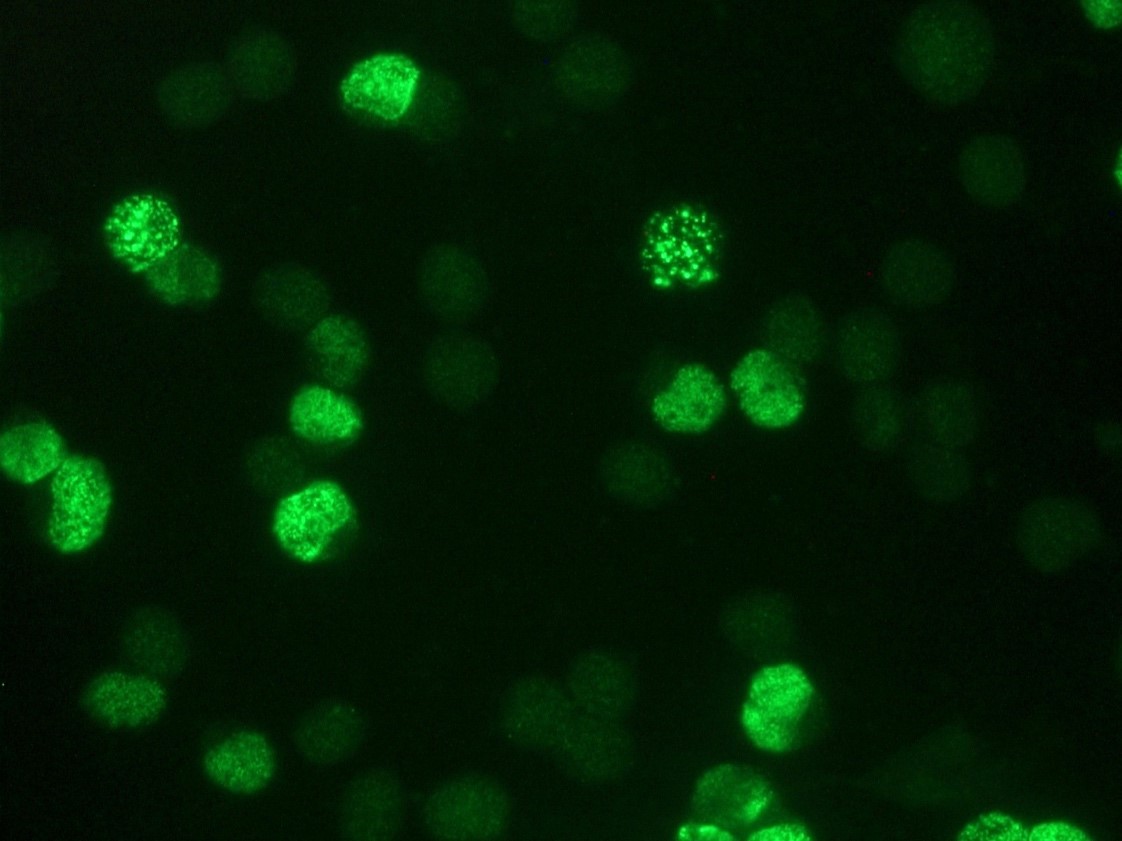
Figure 2: Indirect immunofluorescence staining of BrdU-labeled MR65 lung cancer cells using MUB0200 (IIB5)
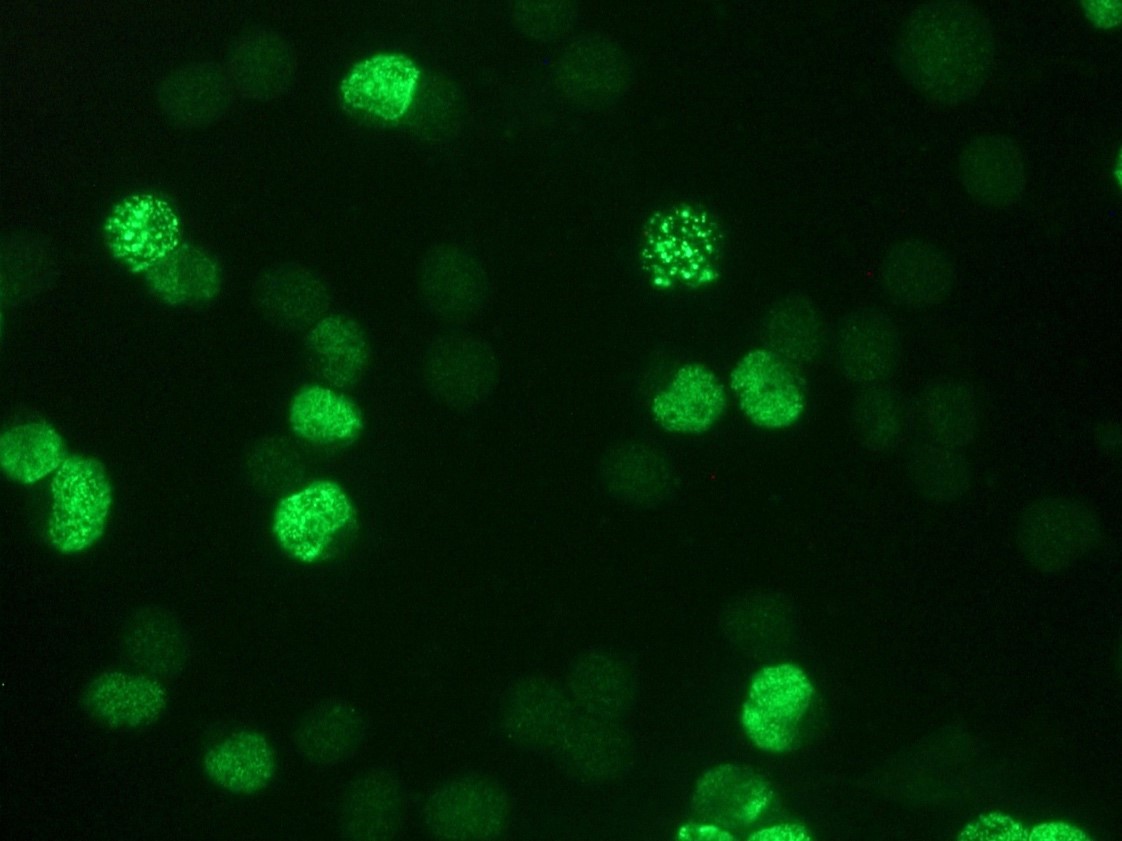
Figure 3: BrdU staining of MR65 human lung cancer cells in culture using MUB0200P (clone IIB5)
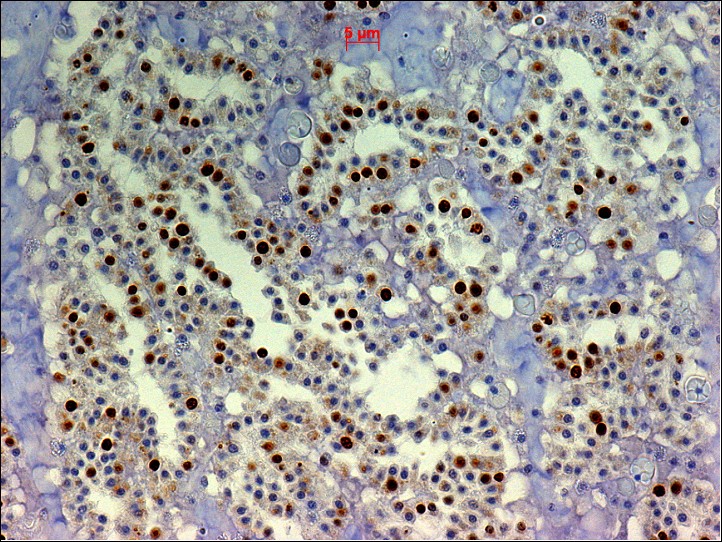
Figure 4: BrdU staining in sponge (Halisarca caerulea) using MUB0200P (clone IIB5)
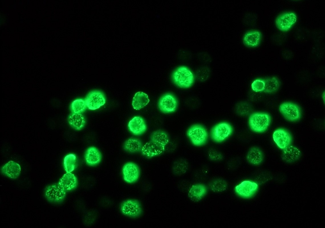
Figure 5. Indirect immunofluorescence staining of BrdU-labeled MR65 lung cancer cells using MUB0200P (IIB5; diluted 1:1000).
